
The Unchanged Room
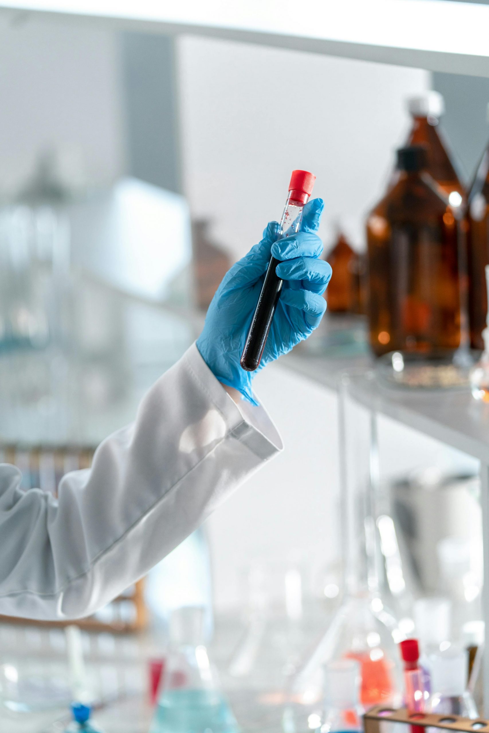
Walk into a molecular biology laboratory today, and you’re stepping into a time capsule. Glass reagent bottles line the shelves. Plastic pipette tips spill from half-empty boxes. Parafilm wrap—that stretchy, waxy film invented in 1933—still seals every container. A pH meter dangles its cord next to a Gilson pipette that would look familiar to a scientist from 1972. The tabletop centrifuge in the corner? Its basic design hasn’t changed in fifty years.
Now walk into an IVF laboratory, and the déjà vu intensifies. The incubators maintaining 37°C and 6% CO₂. The stereomicroscopes with their binocular eyepieces. The micromanipulators for ICSI (intracytoplasmic sperm injection). The culture dishes. The heated stages. Even the meticulous hand-labeling of every dish, every tube, every sample.
If Sydney Brenner—who helped crack the genetic code in the 1960s—could time-travel to 2025 and visit either lab, he’d recognize almost everything. The equipment. The layout. The workflow. The smell of ethanol and agar. He might be confused by the computers, impressed by the resolution of modern microscopes, but fundamentally, he’d know exactly where he was and what to do.
This raises an uncomfortable question: If we can sequence entire genomes for $200, edit DNA with CRISPR, and train AI to predict protein structures, why do our laboratories still look like mid-century museum exhibits?
The answer isn’t stagnation. It’s something more interesting: we got it right the first time.
The Accidental Genius of Lab Design
The modern molecular biology laboratory wasn’t designed by committee or master-planned by engineers. It evolved organically, tool by tool, through decades of trial and error. Each piece of equipment solved a specific problem, and the ones that worked stuck around.
Consider the humble Eppendorf tube—that conical plastic microcentrifuge tube with the snap-cap lid. Why conical instead of flat-bottomed? Because when you spin it in a centrifuge, everything pellets at the point. The cone concentrates your sample into the smallest possible volume, making it easier to see, easier to resuspend, easier to pipette. The snap-cap prevents evaporation but opens with one hand, critical when you’re wearing gloves and holding a pipette.
This wasn’t engineered through sophisticated CAD modeling and fluid dynamics simulations. It was empirical—scientists tried different shapes until they found one that worked brilliantly for almost everything.
The same logic applies to nearly every lab tool:
Petri dishes (invented by Julius Petri in 1887) are shallow cylinders because bacteria need oxygen, and a thin layer of agar maximizes surface area while minimizing diffusion distance. The overlapping lid prevents contamination while allowing gas exchange. Simple. Effective. Unchanged for 138 years.
Erlenmeyer flasks (created by Emil Erlenmeyer in 1860) have their distinctive conical shape because it allows vigorous swirling without spilling—essential for culturing bacteria or cells in liquid media. The narrow neck reduces evaporation. The flat bottom sits stable on hot plates and magnetic stirrers.
Graduated cylinders are tall and narrow because that geometry minimizes the measurement error from meniscus reading. A 100mL volume in a wide beaker is harder to read accurately than the same volume in a narrow cylinder.
Each of these designs is a local optimum—not perfect for every use case, but good enough for most, and the switching costs of replacing them with something marginally better are enormous.
The IVF Laboratory: Where Stakes Get Personal
If molecular biology labs are frozen in time, IVF laboratories are even more conservative—and for good reason. When you’re handling someone’s genetic future, innovation for innovation’s sake becomes reckless.
An IVF lab is a precision environment operating at the edge of what’s biologically possible. Consider what’s actually happening:
Oocyte retrieval extracts immature eggs from follicles, requiring timing precision measured in hours. Too early, and the eggs aren’t mature. Too late, and they ovulate naturally.
Fertilization in vitro demands exact conditions: temperature within 0.3°C of body temperature, CO₂ at 5-6% to maintain pH 7.3-7.4, osmolality at 285-295 mOsm/kg. Deviate from these parameters, and embryos arrest or develop abnormally.
Culture media contain precisely balanced nutrients, amino acids, proteins, and growth factors. The recipes are proprietary and cost thousands of dollars per liter. Every component matters.
Embryo grading at day 3 or day 5 determines which embryos get transferred. Morphological assessment—the symmetry of cells, the appearance of fragmentation, the expansion of the blastocyst—predicts implantation potential. This assessment is still done by trained embryologists looking through microscopes, using criteria established decades ago.
Cryopreservation via vitrification (ultra-rapid freezing) prevents ice crystal formation that would shred cellular membranes. The process requires cooling at rates exceeding 20,000°C per minute, achieved by plunging straws directly into liquid nitrogen at -196°C.
Every step is manual. Every step is critical. Every mistake could cost someone their child.
This is why IVF labs haven’t rushed to automate. The technology works. Success rates have climbed from under 10% per cycle in the 1980s to over 50% for women under 35 today. When the stakes are this high and the process this successful, caution isn’t conservatism—it’s wisdom.
But caution doesn’t mean complacency.
The Problem Hiding in Plain Sight
The current IVF laboratory model has three fundamental vulnerabilities:
1. Human Error at Scale
Every label written by hand is a potential mix-up. Every dish moved between incubators is a potential misidentification. Every embryo graded by visual inspection introduces subjective variability between embryologists.
Witness protection programs don’t prevent mistakes—they limit exposure. Laboratories implement double-witnessing protocols where two embryologists verify every step. But this doubles labor costs and still doesn’t eliminate error. A 2016 study across 28 IVF centers found error rates of 1 in 1,400 cycles—rare, but devastating when it occurs.
The most catastrophic mistakes involve sample swaps: the wrong sperm fertilizing the wrong egg, or the wrong embryo transferred to the wrong patient. These are mercifully rare but represent failures so profound they end careers and shut down clinics.
2. Embryologist Shortage
There aren’t enough trained embryologists to meet demand. Training takes years—typically a PhD in developmental biology or reproductive science, followed by 2-3 years of supervised clinical practice. The work is stressful, the hours irregular (ovulation doesn’t respect 9-to-5 schedules), and burnout is common.
As IVF demand grows—driven by delayed childbearing, increasing infertility rates, and same-sex couples accessing reproductive technology—the shortage worsens. In the US alone, demand is projected to outpace embryologist supply by 40% by 2030.
3. Subjective Variability in Embryo Assessment
Two embryologists looking at the same embryo might grade it differently. One sees “good quality, suitable for transfer.” Another sees “moderate quality, consider waiting for day 5.” These subtle differences in interpretation affect which embryos get transferred, which get frozen, and which get discarded.
Studies show inter-observer agreement rates of only 50-70% for embryo morphology scoring. That means 30-50% of the time, different embryologists would make different decisions about the same embryo.
This isn’t a criticism of embryologists—it’s a limitation of the method. Morphological assessment is subjective, pattern recognition is difficult, and human visual systems aren’t designed to catch subtle gradations in cellular appearance under microscopes.
Enter the Robots (Finally)
For years, automation in IVF meant incremental improvements: better incubators with tighter environmental control, time-lapse imaging systems that photograph embryos every 10 minutes without disturbing them, electronic witnessing systems that use RFID chips to track samples.
These are valuable but limited. They’re assistive technologies, not transformative ones. The core process—human hands manipulating gametes and embryos under microscopes—remained unchanged.
That’s starting to change, and MyBabyBridge represents the vanguard of this transformation.
MyBabyBridge: Robotics Meets Reproductive Medicine
MyBabyBridge is developing robotic systems specifically designed for IVF laboratories, focusing on the tasks where automation offers the greatest benefit: precision, consistency, and error reduction.
Their platform addresses several critical workflows:
Automated embryo culture uses robotic handling to move dishes between incubators, reducing exposure to suboptimal conditions. Every time a human opens an incubator door, the internal environment destabilizes—temperature drops, CO₂ levels fluctuate, humidity changes. Recovery takes minutes. Over five days of culture, these perturbations accumulate. Robots can transfer dishes in seconds, with minimal environmental disruption.
AI-powered embryo selection analyzes time-lapse images using deep learning models trained on thousands of embryo development sequences. The AI doesn’t just look at morphology at one time point—it analyzes the entire developmental trajectory. How quickly did the embryo cleave from 2 cells to 4? Was the timing of compaction normal? Did the blastocyst expand smoothly or irregularly?
These dynamic parameters predict implantation better than static morphology alone. A 2024 study showed AI-assisted selection improved clinical pregnancy rates by 12% compared to traditional morphological grading—equivalent to adding several years of experience to every embryologist.
Automated vitrification standardizes the cryopreservation process. The current method relies on embryologists manually transferring embryos through a series of solutions with increasing cryoprotectant concentrations, then loading them onto specialized devices and plunging into liquid nitrogen. Timing is critical—too long in high-concentration cryoprotectant causes toxicity; too short leaves water in the cells that forms ice crystals.
Robotic systems execute this process with millisecond precision, eliminating timing variability. Early data suggests survival rates post-thaw improve by 3-5%, which may seem small until you remember each percentage point represents actual pregnancies.
The Technical Challenges
Building robots for IVF isn’t trivial. The requirements are extreme:
Sterility: Every surface that touches biological material must be sterile. Robotic grippers, pipette tips, transfer devices—all must either be single-use or autoclavable. Contamination means lost cycles and potential infections.
Precision: Manipulating cells measuring 100-120 micrometers (the size of a human egg) requires positional accuracy within a few micrometers. Robotic actuators must be incredibly precise while also being gentle—too much force and you damage the zona pellucida (the egg’s protective shell).
Speed vs. Gentleness: Moving fast minimizes environmental exposure but increases mechanical stress on cells. The optimal motion profile involves rapid transport followed by extremely gentle deceleration. This requires sophisticated control algorithms.
Integration: IVF labs aren’t empty rooms where you install a robot and start. They’re full of existing equipment—incubators, microscopes, micromanipulators, liquid nitrogen dewars—that cost hundreds of thousands of dollars and can’t be replaced. New robotic systems must integrate with legacy equipment, which means custom adapters, interfaces, and protocols.
Regulatory Approval: Medical devices require FDA clearance or approval, a process that takes years and costs millions. Every component must be validated, every software algorithm verified, every failure mode analyzed. The documentation alone fills rooms.
MyBabyBridge is navigating all of this, building systems that satisfy both engineering requirements and regulatory demands while delivering meaningful clinical benefits.
AI in the Laboratory: Beyond IVF
The transformation happening in IVF labs mirrors broader trends across laboratory medicine and biomedical research. AI and robotics are infiltrating every corner where precision, pattern recognition, and throughput matter.
Pathology: The Digital Microscope Revolution
Histopathology—examining tissue samples under microscopes to diagnose disease—has been fundamentally unchanged since the 1800s. A pathologist receives a slide, places it on a microscope stage, scans it manually, and renders a diagnosis based on cellular morphology, architecture, and staining patterns.
This process is slow (each slide takes 5-10 minutes), subjective (inter-pathologist agreement is 60-80% for many cancers), and limited by human pattern recognition (rare features can be missed in large samples).
Digital pathology is changing this. Whole slide scanners digitize tissue sections at sub-micron resolution, creating gigapixel images. AI algorithms trained on millions of annotated images can detect cancer, grade tumors, identify specific biomarkers, and predict treatment response.
In 2024, FDA-cleared AI systems for prostate cancer detection showed sensitivity exceeding 95%—higher than average pathologists—while reducing false positives by 30%. For breast cancer, AI can identify subtle patterns associated with metastatic potential that human eyes miss.
The technology isn’t replacing pathologists; it’s augmenting them. The AI flags suspicious regions, the pathologist reviews and makes the final call. It’s collaborative intelligence—machine pattern recognition plus human judgment and context.
Radiology: From Film to Algorithms
Radiology was one of the first medical specialties transformed by AI, partly because it was already digital and partly because image analysis is what neural networks do best.
Modern AI systems can:
- Detect lung nodules on CT scans with accuracy matching experienced radiologists
- Identify intracranial hemorrhages on non-contrast head CTs
- Measure tumor volumes and track changes over time
- Predict stroke risk from carotid artery calcification on dental X-rays
- Estimate bone age from hand radiographs for pediatric endocrinology
What’s remarkable isn’t just accuracy—it’s speed. An AI can analyze a chest X-ray in seconds and flag critical findings for immediate attention. In emergency departments with limited radiologist coverage at night, AI triage systems ensure stroke patients and trauma cases get immediate reads while less urgent studies wait until morning.
The economic implications are substantial. Radiologists spend years training to recognize patterns that AI can now learn from labeled datasets in weeks. This doesn’t eliminate the need for radiologists—interpretation still requires clinical context, correlation with patient history, and judgment calls about equivocal findings—but it changes what they spend time on.
Drug Discovery: Robots That Work 24/7
Pharmaceutical research involves mind-numbing amounts of repetitive work. Screening potential drug compounds requires testing thousands to millions of molecules against target proteins, cells, or organisms. Each test involves precise liquid handling—adding reagents, mixing, incubating, measuring fluorescence or luminescence or other readouts.
Humans can perform maybe 100-200 assays per day. High-throughput screening robots can perform 100,000 per day.
Companies like Ginkgo Bioworks operate “foundries”—automated facilities with robots performing the same workflows 24/7 that graduate students used to do by hand. Liquid handlers aspirate and dispense microliter volumes. Plate readers measure 384 samples simultaneously. Robotic arms move plates between stations. The entire process is orchestrated by software.
This isn’t just faster—it’s more consistent. Robots don’t get tired, distracted, or sloppy. Every pipetting action is identical. Every incubation time exact. This reduces experimental noise and improves reproducibility, addressing the replication crisis plaguing biomedical research.
Genomics: The Sequencing Moonshot
DNA sequencing has followed a trajectory steeper than Moore’s Law. The Human Genome Project took 13 years and $3 billion to sequence one genome. Today, a genome costs $200 and takes 24 hours.
This wasn’t just incremental improvement—it was wholesale technology replacement. Multiple times.
Sanger sequencing (first generation) was manual, using radioactive labels and reading sequences from gel electrophoresis. Applied Biosystems automated this with capillary sequencers in the 1990s, turning months of work into hours.
Next-generation sequencing (second generation) parallelized massively—Illumina machines sequence billions of DNA fragments simultaneously by detecting fluorescent nucleotides as polymerase incorporates them. Throughput jumped from megabases to gigabases per run.
Third-generation sequencing (Oxford Nanopore, PacBio) threads single DNA molecules through nanopores or processes them in zero-mode waveguides, reading sequences directly without amplification. Read lengths jumped from 300 bases to 30,000+ bases.
Each generation required new chemistry, new optics, new robotics, and new software. The labs using these machines look nothing like sequencing labs from 20 years ago. Where there were gels and radioactivity, now there are microfluidic chips and laser scanners.
AI entered the picture for base calling—converting raw optical signals into ATCG sequences. Neural networks trained on known sequences learned to distinguish signal from noise, improving accuracy from 99.9% to 99.99%+. That extra nine matters: it’s the difference between 1 error per 1,000 bases and 1 error per 10,000 bases, which determines whether you can reliably detect rare variants.
Surgery: The Robot in the Operating Room
Surgical robots like the da Vinci system have performed millions of procedures since 2000. The robot doesn’t operate autonomously—a surgeon controls it, manipulating instruments through a console while viewing a 3D image of the surgical site.
But the robot provides capabilities human hands lack:
- Motion scaling: The surgeon moves the controls 10 cm, the instruments move 1 cm. This allows microscopic precision.
- Tremor filtering: The robot removes the surgeon’s natural hand tremor.
- Impossible angles: Instruments can rotate 360° and bend in ways human wrists can’t.
- Stable camera: Robotic camera holders don’t get tired or drift.
The result: smaller incisions, less blood loss, faster recovery, fewer complications. For prostate surgery, robotic assistance reduced positive surgical margin rates (cancer cells at the edge of the removed tissue) from 15% to under 10%. For mitral valve repair, robotic approaches reduced ICU stays by 30%.
Now AI is being layered on top. Computer vision systems provide real-time augmented reality overlays—highlighting blood vessels to avoid, outlining tumor margins, showing sub-surface anatomy from pre-operative imaging. Machine learning algorithms trained on thousands of procedures can predict complications before they occur, warning surgeons when instrument movements suggest increased bleeding risk or tissue damage.
The Resistance to Change
Given all these advances, why do so many laboratories—especially core research labs—still look like 1965?
Economic Inertia
Laboratory equipment is expensive and long-lasting. A good centrifuge costs $3,000-15,000 and lasts 20+ years. Microscopes run $10,000-100,000 and become heirlooms passed between graduate students. Replacing functioning equipment with marginally better versions is economically irrational.
This creates a replacement cycle measured in decades. Even when better technology exists, labs don’t upgrade until equipment breaks or becomes truly obsolete.
Training and Muscle Memory
Scientists spend years developing manual skills. Pipetting accurately and precisely is harder than it looks—it requires specific thumb pressure, smooth plunger motion, and timing. Experienced researchers can pipette 5 microliters with better accuracy than cheap electronic pipettes.
When you’ve spent a decade perfecting these skills, automation feels like admitting your expertise is obsolete. There’s psychological resistance to technologies that make your hard-won abilities irrelevant.
The “If It Ain’t Broke” Mentality
Science is conservative by nature. You don’t get published by reproducing known results with fancy new equipment. You get published by discovering something new, and the safest path is using proven methods.
Early adopters of new technology risk failure modes that aren’t their fault—bugs in software, quirks in automation, unexpected interactions. These delays can kill projects, ruin careers, and waste grants. Better to stick with methods that work, even if they’re tedious.
Regulatory Barriers (For Clinical Labs)
Clinical laboratories face additional hurdles. Every assay must be validated, every instrument qualified, every procedure documented. Introducing new technology requires revalidation, which means weeks of running samples in parallel on old and new systems, demonstrating equivalence, and documenting everything for regulatory inspectors.
The paperwork alone discourages innovation. Labs stick with “grandfathered” methods that predate modern regulatory requirements rather than adopt newer, better ones that trigger full validation requirements.
Why Change Is Accelerating Now
Despite these barriers, laboratory automation is accelerating, driven by converging forces:
1. The AI Inflection Point
Machine learning works when you have:
- Large datasets (genomics, medical imaging, lab data)
- Clear objectives (diagnose disease, predict outcomes, identify patterns)
- Measurable performance (accuracy, sensitivity, specificity)
All three are now abundant in laboratory medicine. Medical data generation has exploded. Computational power is cheap. Pre-trained models can be fine-tuned for specific tasks with modest datasets.
The result: AI applications that actually work, not science fiction. They deliver measurable improvements in accuracy, speed, or cost. This creates economic pressure to adopt.
2. The Talent Shortage
There aren’t enough trained professionals—embryologists, pathologists, cytotechnologists, clinical laboratory scientists—to meet demand. Automation becomes necessary, not optional.
Healthcare systems facing technologist shortages have three options: pay higher salaries (expensive), train more people (slow), or automate (practical). Increasingly, they’re choosing the third.
3. Precision Medicine Requires Throughput
Modern medicine is moving toward personalized treatment based on individual genetics, biomarkers, and disease characteristics. This requires massive testing—sequencing tumor DNA, profiling immune cells, measuring dozens of proteins simultaneously.
Manual methods can’t scale. A pathologist can review 50 slides per day. A precision oncology case might require analyzing mutations in 500 genes, expression levels of 20,000 genes, and immune infiltration patterns across dozens of tissue regions. That’s not humanly feasible without computational assistance.
4. Consumer Expectations
Patients increasingly expect precision, speed, and transparency. When they can track a pizza delivery in real-time, they wonder why embryo culture is a black box. When AI can diagnose skin cancer from phone photos, they question why their biopsy takes two weeks.
Consumer pressure drives clinics to adopt technologies that provide better information, faster results, and more certainty. IVF clinics advertising “AI-powered embryo selection” attract patients. Pathology groups offering 24-hour turnaround times win market share.
The Laboratory of 2040
What will laboratories look like in fifteen years?
Smaller, Smarter, More Integrated
Current labs are sprawling spaces packed with equipment. Future labs will be compact, with modular automation replacing rows of individual instruments.
Imagine a single “synthesis and analysis core”—a refrigerator-sized unit that performs sample preparation, genomic sequencing, proteomics, and metabolomics. Microfluidic chips replace racks of tubes. Nanoliter reactions replace milliliter volumes. AI-driven quality control identifies problems before experiments fail.
IVF labs might shrink to the size of a small office, with automated culture systems, integrated time-lapse imaging, AI embryo selection, and robotic vitrification all in one unit. The embryologist becomes a quality controller and decision-maker rather than manual operator.
AI-First Workflows
Every experimental result will be immediately analyzed by AI:
- Sequence quality assessment
- Contamination detection
- Statistical analysis
- Literature searches for similar findings
- Suggested follow-up experiments
Scientists won’t wait weeks for results and then spend days analyzing them. They’ll have preliminary insights while experiments run, allowing real-time course corrections.
In clinical labs, AI will triage every sample:
- Flag critical results immediately
- Route routine results to automated reporting
- Send equivocal cases to human experts with relevant context highlighted
Distributed and Decentralized
Not all testing will require centralized laboratories. Point-of-care devices will bring sophisticated diagnostics to clinics, pharmacies, and homes.
We’re already seeing this:
- Blood glucose monitors for diabetics
- INR testing for patients on warfarin
- COVID tests at home
- Continuous glucose monitors sending data to smartphones
Future generations will be more capable:
- Rapid PCR for infection diagnosis at urgent care clinics
- Portable mass spectrometers for drug monitoring
- Handheld ultrasound with AI interpretation
- Wearable sensors monitoring dozens of biomarkers continuously
Some IVF procedures might move closer to patients. Embryo culture might happen in small, automated systems at local clinics rather than requiring travel to major fertility centers. Gamete processing and initial fertilization could be distributed, with only complex cases requiring specialist facilities.
Human-AI Collaboration as Standard
The future isn’t human vs. machine—it’s human plus machine. Embryologists will use AI to shortlist embryos, then make final decisions considering patient history, prior cycles, and preferences. Pathologists will review AI-flagged regions rather than scanning entire slides. Surgeons will operate with real-time AI guidance about anatomy and risk.
This collaboration plays to each side’s strengths:
- AI: Pattern recognition, consistency, tirelessness, speed
- Humans: Context, judgment, creativity, empathy
The best outcomes come from combining both.
The Global Health Congress Conversation
These transformations—robotics in IVF, AI in pathology and radiology, automation in drug discovery—represent converging revolutions in how we practice medicine and conduct research. They’re happening simultaneously across specialties but often in isolation, with limited cross-pollination.
An embryologist working with MyBabyBridge’s robotic systems might have valuable insights for the surgical robotics team, but they rarely occupy the same conferences. A pathologist using AI for cancer diagnosis could inform drug discovery scientists about automated image analysis, but those communities don’t naturally overlap. A genomics researcher developing new sequencing protocols might benefit from IVF lab sterility procedures, but who makes that connection?
This is precisely why GlobalHealthCongress.com is bringing together experts from across these domains. We’re creating dedicated sessions where:
IVF specialists meet surgical roboticists to compare approaches to precision manipulation of biological materials
Pathologists using AI discuss with radiologists using AI to identify transferable algorithms and shared challenges in medical imaging analysis
Drug discovery automation engineers exchange notes with clinical laboratory automation specialists about liquid handling precision, sample tracking, and error prevention
Embryologists, genetic counselors, and precision medicine teams explore how AI-assisted decision-making can improve outcomes while maintaining human judgment and ethical oversight
Medical device developers, regulatory experts, and clinicians work through the challenges of bringing innovative technologies from prototype to clinical practice
The goal isn’t just knowledge sharing—it’s catalyst creation. When the person developing robotic grippers for embryo manipulation meets the person designing robotic suturing systems for surgery, they realize they’re solving the same force-sensing problem with different tools. That conversation might spark a collaboration that accelerates both fields.
When the AI researcher training models on embryo morphology meets the AI researcher training models on tumor histology, they discover their datasets have similar challenges—limited examples of rare but important categories, subjective ground truth labels, and need for explainable predictions. That shared understanding might lead to new techniques benefiting both.
The Convergence Ahead
The laboratory of 1965 emerged organically, one tool at a time, through empirical refinement. It worked brilliantly for decades because those tools solved fundamental problems elegantly.
The laboratory of 2040 will emerge intentionally, designed around capabilities rather than legacy constraints. It will combine:
- The precision of robotics
- The pattern recognition of AI
- The speed of automation
- The judgment of human experts
- The adaptability of modular systems
We’re living through the transition. MyBabyBridge in IVF labs. AI in pathology and radiology. Robotic surgery. Automated drug discovery. Each represents a thread in a larger transformation of how we practice medicine and conduct research.
The question isn’t whether this transformation will happen—it’s already underway. The question is how quickly we can learn from each other, transfer insights across domains, and accelerate the adoption of technologies that improve outcomes.
That requires conversation. Sustained, cross-disciplinary, technically deep conversation between people who’ve spent careers in different corners of laboratory medicine and biomedical research.
That’s what we’re building.
Join us at GlobalHealthCongress.com for in-depth sessions on laboratory automation, AI in medicine, robotic systems in clinical care, and the future of precision healthcare. Featuring demonstrations from innovators like MyBabyBridge, technical workshops on AI implementation, and collaborative working groups addressing the challenges of bringing cutting-edge technology into clinical practice. Because the future of the laboratory won’t be designed in isolation—it will emerge from conversations between experts who bring different pieces of the puzzle.
I am Founder and CEO of Mybabybridge and in Leadership Team of Global Health Forum.
Further Reading
On IVF Laboratory Technology:
- Meseguer, M., et al. (2024). “Time-lapse imaging and artificial intelligence in embryo selection.” Fertility and Sterility, 121(3), 421-433.
- Chavez-Badiola, A., et al. (2023). “Automated embryo culture and selection systems.” Human Reproduction, 38(6), 1156-1167.
On AI in Medicine:
- Topol, E. (2024). Deep Medicine: How Artificial Intelligence Can Make Healthcare Human Again (2nd ed.). Basic Books.
- Esteva, A., et al. (2023). “A guide to deep learning in healthcare and the life sciences.” Nature Medicine, 29, 1871-1888.
On Laboratory Automation:
- King, R.D., et al. (2024). “The automated laboratory: From inception to implementation.” Nature Reviews Methods Primers, 4, 18.
- Yachie-Kinoshita, A., et al. (2023). “Modeling the future of biological laboratories.” Trends in Biotechnology, 41(9), 1234-1247.
On Surgical Robotics:
- Sheetz, K.H., et al. (2024). “Evolution of robotic surgery: Past, present, and future.” Annals of Surgery, 279(1), 45-54.
- Alemzadeh, H., et al. (2023). “AI-augmented robotic surgery: Opportunities and challenges.” IEEE Transactions on Medical Robotics, 5(2), 178-192.
