
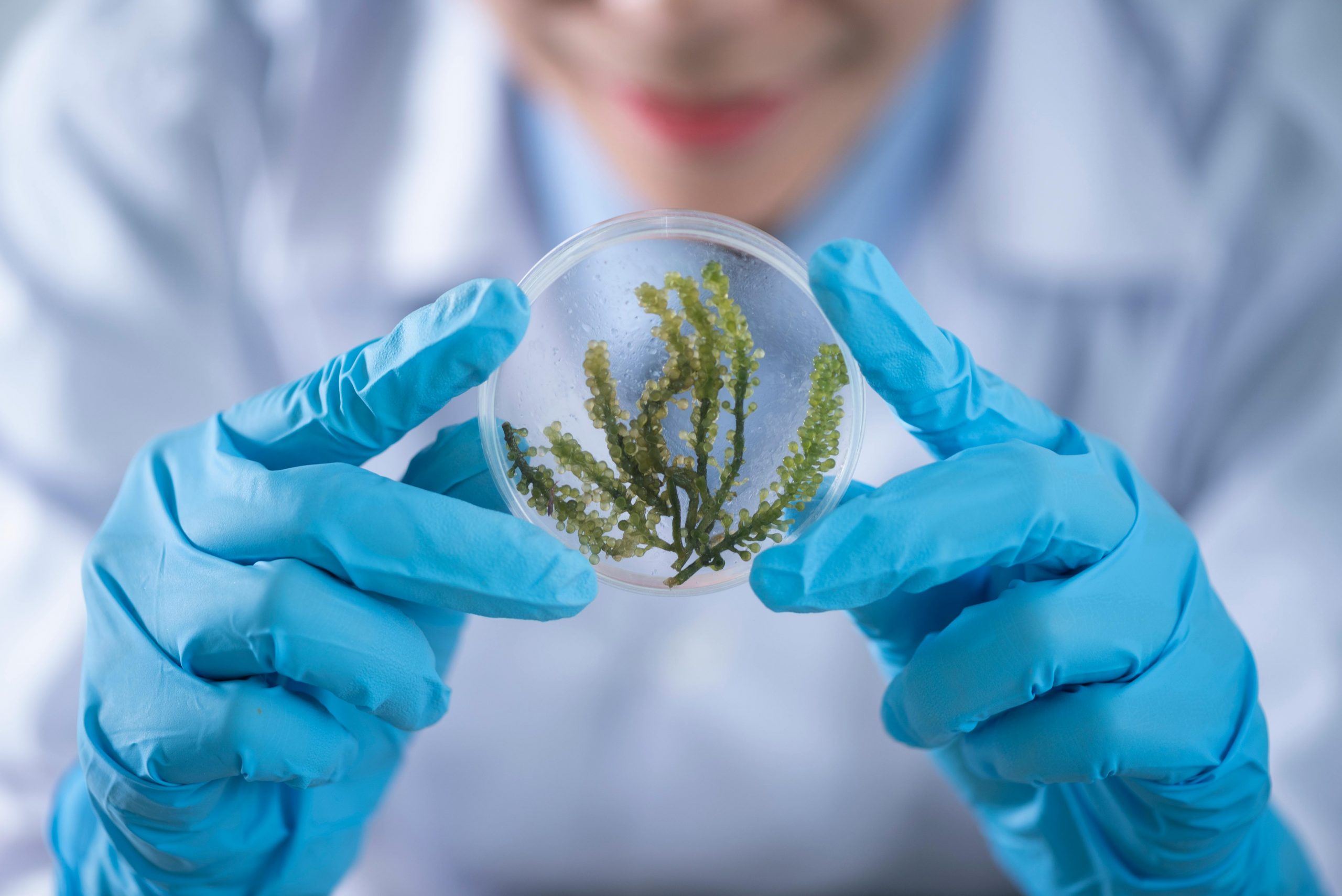
The Green Fluorescent Protein Problem
In 1994, Martin Chalfie shocked the scientific world by making E. coli bacteria glow green. He’d inserted a single gene from the jellyfish Aequorea victoria—encoding green fluorescent protein (GFP)—and suddenly, living cells became visible under blue light. For this breakthrough, he shared the 2008 Nobel Prize in Chemistry.
GFP revolutionized biology. Before GFP, tracking proteins inside cells required killing the cells, fixing them, and staining them with antibodies—a snapshot of death. GFP enabled live imaging. Scientists could watch proteins move in real-time, see when genes turned on or off, and observe biological processes as they happened.
Today, GFP is everywhere. Nearly every molecular biology lab uses fluorescent proteins to visualize cellular processes. Researchers have engineered variants spanning the entire visible spectrum—cyan, yellow, red, far-red. There are photoactivatable versions that turn on when hit with specific wavelengths. pH-sensitive versions that change color based on acidity. Versions optimized for brightness, stability, or speed of maturation.
But GFP has fundamental limitations that restrict its utility beyond the laboratory bench:
1. It requires external illumination. You need to shine blue light on cells to make them fluoresce. Inside opaque tissues—like tumors, organs, or soil—you can’t get light in and you can’t see the fluorescence out.
2. Visible light doesn’t penetrate tissue. Light in the visible spectrum (400-700 nm) travels only about 1 millimeter into tissue before being scattered and absorbed. This is why you can’t see through your hand even with a bright flashlight.
3. It produces weak signals. Fluorescence is inherently dim—each fluorescent molecule emits far fewer photons than it absorbs. Detecting GFP requires sensitive cameras, dark rooms, and close proximity.
4. It’s drowned out by background autofluorescence. Many biological molecules naturally fluoresce—NADH, flavins, collagen, elastin. This background noise makes detecting GFP-labeled cells in complex environments like soil, water, or living tissue extremely difficult.
These limitations mean GFP works beautifully in controlled laboratory settings but fails in almost every real-world application. You can’t use it to monitor bacteria in agricultural fields, detect pathogens in water supplies, track engineered microbes in bioreactors, or image tumors deep inside bodies.
For biology to move from lab benches to real-world applications—environmental monitoring, disease diagnostics, industrial bioprocessing—we need fundamentally different ways for cells to communicate what they’re sensing.
The Transducer Bottleneck
Every biosensor has two components:
The sensor detects a target—a chemical, temperature change, mechanical force, or biological molecule. Nature evolved countless sensors: receptors that bind hormones, ion channels that respond to voltage, olfactory receptors that detect odor molecules, photoreceptors that sense light.
The transducer converts that detection event into a measurable signal. In electronics, transducers convert physical phenomena (pressure, temperature, light) into electrical signals. In biology, transducers convert molecular recognition into observable outputs—fluorescence, color change, electrical signals.
Bioengineers have gotten excellent at borrowing sensors from nature. Want to detect TNT? Use receptors from bacteria that metabolize nitroaromatic compounds. Need to sense glucose? Adapt bacterial glucose-binding proteins. Looking for specific DNA sequences? CRISPR-Cas systems naturally recognize target sequences.
But we’ve been terrible at developing diverse transducers. Almost every biosensor developed in the last 30 years uses one of three outputs:
- Fluorescent proteins (GFP, RFP, mCherry, etc.)
- Luciferases (enzymes that produce bioluminescence)
- Colorimetric reporters (pigments visible to the naked eye)
All three share the same fundamental problem: they rely on visible light, which doesn’t travel through tissues, soil, water, or air at useful distances.
This transducer bottleneck limits biosensor applications. We can engineer bacteria to detect explosives, but we need to grow them on agar plates and shine UV light to see the response. We can create cells that sense tumor markers, but we can’t implant them and monitor them non-invasively. We can design microbes that detect water pollution, but we need to collect samples, concentrate cells, and examine them under microscopes.
The transducer bottleneck is why biosensors remain mostly research tools rather than deployed technologies.
Breaking Through: New Ways to Listen to Cells
Recent innovations are finally expanding our transducer toolkit, enabling biosensors to work in previously inaccessible environments.
Ultrasound-Based Reporters
Ultrasound penetrates tissue far better than light—it’s how we image fetuses, hearts, and tumors non-invasively. In 2018, Mikhail Shapiro’s lab at Caltech engineered the first genetically encoded ultrasound reporters using gas vesicles.
Gas vesicles are protein nanostructures that bacteria and archaea naturally produce to control buoyancy in aquatic environments. They’re hollow protein shells filled with gas, measuring about 250 nanometers. When hit with ultrasound waves, they scatter sound differently than surrounding tissue, creating contrast in ultrasound images.
Shapiro’s team identified the genes encoding gas vesicles, transferred them into mammalian cells, and demonstrated that these cells become visible on standard clinical ultrasound machines. Crucially, gas vesicle expression can be linked to biosensor circuits—the cells only produce gas vesicles when they detect their target molecule.
Applications:
Imaging engineered cells in vivo: Inject bacteria engineered to colonize tumors and produce gas vesicles when they detect low oxygen (a tumor marker). Track tumor location and oxygenation status non-invasively with ultrasound.
Monitoring cell therapy: CAR-T cells engineered to produce gas vesicles when activated can be tracked as they find and attack cancer cells.
Industrial bioprocess monitoring: Bacteria in large bioreactors can report their metabolic state acoustically, enabling real-time monitoring without sampling.
The technology is advancing rapidly. By 2023, researchers had engineered gas vesicles with different shapes and sizes that scatter ultrasound at distinct frequencies, creating a “acoustic color palette” analogous to fluorescent proteins of different colors.
MRI-Based Reporters
Magnetic resonance imaging (MRI) provides exquisite anatomical detail anywhere in the body. In 2018, Alan Jasanoff’s lab at MIT developed genetically encoded MRI contrast agents.
MRI detects hydrogen atoms (mostly in water) and their interactions with surrounding molecules. Contrast agents—typically gadolinium-based chemicals injected intravenously—alter local magnetic fields, making specific tissues brighter or darker.
Jasanoff’s team discovered that certain bacterial proteins, when expressed in cells, create local magnetic field disturbances detectable by MRI. The key protein, ferritin, naturally stores iron. When overexpressed, cells accumulate iron in ferritin cages, creating magnetic susceptibility differences visible on MRI scans.
By linking ferritin expression to biosensor circuits, cells report their sensing state via MRI. Detect inflammation? Express ferritin. See the inflammation on MRI scans without injecting contrast agents.
This enables:
Functional brain imaging with cellular resolution: Engineer neurons to produce ferritin when they fire. Map brain activity with spatial resolution and molecular specificity impossible with fMRI alone.
Tracking immune responses: T cells engineered to express ferritin when activated reveal where and when immune responses occur inside tumors or at sites of infection.
Non-invasive glucose monitoring: Engineer cells implanted subcutaneously to produce ferritin in response to glucose levels. Diabetics could monitor blood sugar via quick MRI scans instead of finger pricks.
Hyperspectral Detection from Above
The most radical transducer innovation: making bacteria visible from 90 meters in the air using drones.
This work, published in Nature Biotechnology by Chris Voigt’s lab at MIT, uses hyperspectral imaging—cameras that capture hundreds of narrow wavelength bands instead of just red, green, and blue.
Every molecule absorbs and reflects light in a unique pattern—its spectral fingerprint. Chlorophyll absorbs blue and red but reflects green (why plants look green). Carotenoids absorb blue-green light. Each pigment creates characteristic peaks and troughs in its absorption spectrum.
Hyperspectral cameras were developed by NASA in the 1980s for remote sensing—identifying minerals from satellites, mapping ocean algal blooms, assessing crop health from aircraft. The military uses them to distinguish real rocks from plastic-cased explosives disguised as rocks.
Voigt’s team asked: if hyperspectral imaging can distinguish plastic from rock, why not bacteria from soil?
The challenge: finding molecules that bacteria can make that produce distinctive spectral signatures visible against background noise of soil, plants, and dirt.
They screened thousands of metabolites computationally, using quantum chemistry simulations to predict absorption spectra. Most biological molecules absorb in crowded regions of the spectrum where soil and plants also absorb. But a few absorb in “quiet” wavelength windows—particularly near-infrared (700-1000 nm)—where biological backgrounds are minimal.
They identified two candidates:
Biliverdin IXα: A green pigment produced when heme (from hemoglobin) breaks down. They engineered Pseudomonas putida to produce large quantities by adding two enzymes.
Bacteriochlorophyll a: A photosynthetic pigment from purple bacteria that absorbs infrared light at 860 nm. They engineered Rhodocyclus gelatinosus, which naturally makes small amounts, to produce it in much higher concentrations.
They sprayed these engineered bacteria onto soil at Fort Devens military facility and flew a hyperspectral drone overhead at 90 meters altitude. The algorithm detected bacterial colonies clearly, distinguishing their spectral signatures from surrounding soil, plants, and debris.
This opens extraordinary applications:
Agricultural monitoring: Engineer bacteria that colonize plant roots and produce hyperspectral reporters when they detect nutrient deficiencies, pathogens, or soil contamination. Farmers fly drones over fields, identifying problem areas before visible symptoms appear.
Environmental surveillance: Deploy bacteria that sense explosives, landmines, or chemical weapons and signal their location via hyperspectral signatures visible from aircraft or satellites.
Ecosystem monitoring: Release bacteria engineered to detect pollutants—heavy metals, pesticides, industrial contaminants—in rivers, forests, or wetlands. Monitor environmental health remotely across entire landscapes.
Disaster response: After oil spills or chemical accidents, deploy bacteria that sense and report contamination extent, guiding cleanup efforts.
The bacteria become living sensors, broadcasting their findings into space for anyone with the right camera to see.
The Medical Diagnostics Connection
These transducer innovations aren’t just for environmental monitoring—they’re transforming medical diagnostics and therapeutic monitoring.
Circulating Tumor DNA Detection
Current cancer screening methods—mammograms, colonoscopies, PSA tests—are either invasive, insensitive, or both. Detecting cancer early, when it’s most treatable, requires finding molecular signs before tumors grow large.
Circulating tumor DNA (ctDNA)—fragments of tumor DNA shed into bloodstream—offers a potential solution. Even small tumors release DNA, and detecting tumor-specific mutations in blood samples can identify cancer months or years before traditional screening.
But ctDNA is incredibly rare: one tumor DNA molecule among 10,000 normal DNA molecules. Current detection methods use PCR or sequencing, which are expensive, require laboratory processing, and take days to weeks for results.
Biosensor-based detection could change this. Engineer bacteria carrying CRISPR systems that recognize tumor-specific mutations. When they bind target ctDNA, they activate transducer expression—gas vesicles for ultrasound detection, ferritin for MRI, or secreted biomarkers detectable in urine.
Inject these bacteria intravenously. They circulate, sampling blood for ctDNA. When they find tumor DNA, they produce their reporter. Ultrasound scans reveal where tumor-sensing bacteria accumulated. Blood or urine tests detect secreted biomarkers.
This approach could enable:
- Liquid biopsies for early cancer detection
- Real-time monitoring of treatment response
- Detection of minimal residual disease after surgery
- Personalized screening based on individual genetic risk
Implantable Diagnostic Devices
Instead of drawing blood for lab testing, imagine implanting engineered cells that continuously monitor biomarkers and report wirelessly.
Encapsulate engineered bacteria or mammalian cells in biocompatible hydrogels. The hydrogel allows small molecules (glucose, hormones, inflammatory markers) to diffuse in but protects cells from immune attack. Link biosensors to MRI-visible reporters.
Implant subcutaneously. The cells continuously sample interstitial fluid, responding to biomarker concentrations. Periodic MRI scans—potentially at-home devices for chronic disease management—reveal biomarker levels based on reporter signal intensity.
Applications:
Diabetes management: Glucose-sensing cells eliminate finger-stick testing. Quick MRI scans provide continuous glucose monitoring without wearable devices that can fail or cause skin irritation.
Cardiovascular disease: Cells sensing inflammatory markers, cardiac troponins, or natriuretic peptides (indicators of heart failure) provide early warning of deteriorating cardiac function.
Kidney disease: Monitor creatinine, urea, and electrolytes continuously, detecting acute kidney injury before symptoms appear.
Therapeutic drug monitoring: Cells detect drug concentrations, ensuring patients maintain therapeutic levels without over- or under-dosing.
Transplant rejection surveillance: Implanted cells near transplanted organs detect immune activation markers, catching rejection early when it’s reversible.
Point-of-Care Infectious Disease Testing
Current infectious disease diagnostics require either:
- Culturing organisms (days to weeks)
- PCR testing (hours, requires lab infrastructure)
- Antibody tests (miss early infections)
Biosensor-based detection could enable rapid, sensitive, point-of-care testing.
Engineer bacteria with sensors for pathogen-specific molecules—viral proteins, bacterial cell wall components, parasite antigens. Link to colorimetric or luminescent reporters visible without equipment.
Add patient sample (saliva, blood, urine) to freeze-dried bacteria on paper. If pathogens are present, bacteria activate and produce visible signal within minutes to hours.
This approach offers:
- Equipment-free testing deployable anywhere
- Sensitivity approaching PCR (bacteria amplify signals)
- Multiplexed detection (different bacteria sense different pathogens simultaneously)
- Low cost (bacterial reagents are cheap to produce and store)
Especially valuable in resource-limited settings where lab infrastructure is absent but infectious disease burden is highest.
The AI-Enhanced Sensing Future
Biosensors generate data—lots of it. Interpreting that data, extracting meaningful signals from noise, and predicting outcomes requires computational sophistication that AI provides.
Hyperspectral Image Analysis
The Fort Devens experiment generated terabytes of hyperspectral data—hundreds of wavelength bands for millions of pixels. Finding bacterial signatures buried in complex spectral backgrounds requires algorithms that can:
- Separate signal from noise across 100+ wavelength dimensions
- Account for variable lighting conditions, atmospheric interference, and seasonal changes in background vegetation
- Distinguish target bacteria from naturally occurring organisms with similar spectral properties
- Integrate spatial patterns (bacteria cluster in specific locations) with spectral patterns
Machine learning excels at this. Train convolutional neural networks on labeled hyperspectral images (pixels containing bacteria vs. background). The network learns which spectral features and spatial patterns indicate bacterial presence.
As more hyperspectral biosensor data accumulates, models improve. They’ll detect bacteria at lower concentrations, through partial plant cover, in varied soil types, under different weather conditions.
Eventually, AI might identify bacterial sensing states not just from engineered reporters, but from subtle changes in native bacterial metabolism—detecting stressed bacteria, nutrient-limited bacteria, or bacteria responding to environmental toxins even without genetic engineering.
Multi-Modal Sensor Fusion
Real-world diagnostics often combine multiple sensing modalities. A cancer diagnostic might integrate:
- ctDNA detection via implanted bacteria
- Protein biomarkers in blood
- Imaging findings (CT, MRI, ultrasound)
- Clinical symptoms and patient history
AI can integrate these heterogeneous data streams, weighting each according to reliability and extracting patterns no single test reveals.
For example, slightly elevated ctDNA alone might be inconclusive. But combined with ultrasound showing gas vesicle-expressing bacteria accumulated in a specific organ, inflammatory markers elevated in blood work, and recent onset of symptoms, AI classifies this as high probability early-stage cancer requiring immediate biopsy.
Google’s DeepMind published research in 2024 showing AI models integrating genomic, proteomic, metabolomic, and imaging data predicted cancer outcomes better than any single data type. As biosensor technologies mature, they’ll feed into these multi-modal diagnostic AI systems.
Predictive Microbiology
In environmental monitoring, detecting problems after they occur is reactive. Predicting problems before they manifest is transformative.
AI trained on historical biosensor data—bacterial stress responses in agricultural fields, pollution patterns in watersheds, pathogen detection frequencies in food processing facilities—can predict future issues.
If biosensors detect early signs of fungal infection in wheat fields (specific volatile organic compounds, changes in soil microbiome composition), AI trained on years of crop disease data predicts likelihood of full infection spread, optimal intervention timing, and expected yield impact under different treatment scenarios.
In clinical settings, biosensors monitoring implanted cells could detect patterns preceding disease exacerbations. AI might identify that certain biomarker fluctuation patterns precede heart failure episodes by 72 hours, enabling preventive interventions before patients need emergency hospitalization.
The Robotics and Automation Imperative
Deploying biosensors at scale—monitoring ecosystems, screening populations, operating commercial bioprocesses—requires automation that doesn’t exist yet.
Automated Sample Collection and Processing
Current biosensor workflows involve manual steps:
- Collecting environmental samples (soil, water, air)
- Preparing samples (diluting, filtering, concentrating)
- Applying samples to biosensor cells
- Incubating
- Imaging or measuring output
- Recording and analyzing data
For laboratory research on dozens of samples, this is manageable. For industrial applications requiring thousands to millions of measurements, it’s impossible.
Automated systems must:
Collect samples autonomously: Drones land, collect soil samples, return to base stations. Underwater robots sample rivers and oceans. Stationary monitoring stations continuously draw air or water samples.
Prepare samples without human intervention: Robotic liquid handlers dilute, filter, and aliquot samples into multiwell plates containing freeze-dried biosensor bacteria. Each well gets precisely measured sample volumes.
Incubate under controlled conditions: Automated incubators maintain optimal temperature, humidity, and atmosphere for biosensor activation.
Image or measure outputs automatically: High-content imaging systems photograph all wells. Spectrophotometers measure absorbance. Luminometers detect bioluminescence. Ultrasound arrays scan for gas vesicle signals.
Analyze data in real-time with AI: Results feed directly into machine learning pipelines that classify samples, flag anomalies, and trigger alerts when biosensors detect targets.
Resupply consumables autonomously: When reagents run low, systems order replacements. When biosensor bacteria lose viability, systems prepare fresh cultures.
Companies developing autonomous laboratory systems—like Emerald Cloud Lab, which operates remote-access labs where users command robots via software—point toward this future. But adapting these systems for field deployment in agriculture, environmental monitoring, or disaster response remains challenging.
Bioreactor Integration
Industrial biotechnology uses engineered microbes to produce pharmaceuticals, chemicals, and fuels in bioreactors. Monitoring these processes requires frequent sampling and offline analysis—slowing response times and increasing contamination risk.
Integrating biosensors directly into bioreactors enables real-time monitoring:
Metabolic state sensors: Engineer production bacteria to express ultrasound-visible reporters when stressed, nutrient-limited, or producing product optimally. Ultrasound probes mounted in bioreactor walls provide continuous monitoring without sampling.
Contamination detection: Include sentinel bacteria that detect and report presence of contaminating organisms. Early detection enables intervention before entire batches are lost.
Product titer monitoring: Link biosensors to product concentration, providing real-time measurements that guide harvest timing and downstream processing.
Robotics handle sample collection from automated sample ports, transfer to biosensor chambers, incubation, measurement, and data logging. Facility operators monitor dashboards showing real-time biosensor outputs across all bioreactors, intervening only when AI flags anomalies.
The Regulatory Challenge
Deploying engineered bacteria into open environments triggers intense regulatory scrutiny—justifiably. Once released, microbes can spread, evolve, and interact with ecosystems in unpredictable ways.
Current US regulations classify engineered microbes based on engineering method rather than safety profile. Any organism containing genes from another genus gets flagged for extensive review under EPA’s Toxic Substances Control Act (TSCA). Between 1987 and 2018, over 200 TSCA submissions were filed. None led to commercialized products.
This regulatory structure made sense in 1987 when genetic engineering was new and risks uncertain. Four decades later, with vastly more knowledge about engineered microbes’ environmental fate and impact, the rules are obsolete.
Modernization Approaches
Several strategies could enable safer, faster deployment:
Risk-based assessment: Evaluate organisms based on actual hazard (pathogenicity, persistence, gene transfer potential) rather than modification method. Non-pathogenic bacteria with engineered reporters that don’t confer fitness advantages pose minimal risk.
Containment systems: Engineer obligate dependencies—bacteria that require synthetic nutrients unavailable in nature, or that express kill switches when they leave target environments. These “biological containment” strategies prevent uncontrolled spread.
Genome minimization: Use microbes with reduced genomes lacking genes for horizontal gene transfer, antibiotic resistance, or virulence. These organisms are less likely to persist outside controlled settings or exchange DNA with wild microbes.
Gradual deployment: Start with closed-system applications (bioreactors, contained facilities), prove safety and efficacy, then expand to semi-open systems (agricultural fields with monitoring), and finally fully open environments only after extensive safety data accumulation.
International harmonization: Coordinate regulations across countries to enable technology transfer and avoid regulatory arbitrage where companies deploy in regions with weaker oversight.
Several companies are pioneering pragmatic approaches:
Pivot Bio engineered nitrogen-fixing bacteria for agriculture by remodeling existing genomes without cross-genus gene transfer, simplifying regulatory approval.
Fieldstone Bio (spun out from Voigt’s lab) is developing hyperspectral biosensors using bacteria that don’t contain foreign DNA, potentially avoiding TSCA requirements.
Ginkgo Bioworks partners with regulators early in development, providing extensive safety data and environmental monitoring plans that build confidence.
As successful deployments accumulate safety records—thousands of acres treated, millions of measurements collected, years of environmental monitoring—regulatory agencies will gain evidence supporting streamlined approval for low-risk applications.
The Global Health Congress Intersection
The technologies converging to enable widespread biosensor deployment—engineered microbes, novel transducers, AI analysis, robotic automation, regulatory reform—represent threads of a larger transformation in how we monitor health and environment at population scale.
But these fields evolve in parallel with limited cross-talk:
Synthetic biologists engineering hyperspectral reporters rarely connect with remote sensing specialists optimizing drone imaging systems.
Medical diagnostics developers creating ctDNA biosensors don’t usually collaborate with agricultural microbiologists deploying soil monitoring systems, despite using similar transducer technologies.
AI researchers training models on hyperspectral data don’t typically engage with clinical informaticists integrating multi-modal diagnostic data, though both face similar signal extraction challenges.
Bioprocess engineers instrumenting bioreactors with biosensors operate separately from IVF lab directors monitoring embryo culture systems (like MyBabyBridge), despite shared needs for sterile automated sensing in living systems.
Regulatory experts navigating EPA approval for environmental releases don’t routinely discuss strategies with those pursuing FDA approval for implanted diagnostic cells, missing opportunities to harmonize approaches.
This is what GlobalHealthCongress.com addresses directly. We’re creating forums where:
Transducer innovators from academic labs meet diagnostic device companies to accelerate translation of research tools into commercial products
Hyperspectral imaging specialists work with pathologists to explore whether spectral analysis techniques detecting bacteria from drones can improve tissue diagnostics under microscopes
Robotics engineers from agricultural monitoring systems exchange insights with IVF automation developers (like MyBabyBridge teams) about handling biological materials precisely in variable conditions
Regulatory strategists from environmental and medical sectors compare approval pathways, identifying opportunities for harmonization and best practices transferable across applications
AI scientists working on biosensor data interpretation collaborate with clinicians to ensure models produce actionable, clinically meaningful outputs rather than just statistical associations
The goal isn’t just knowledge sharing—it’s solving shared technical challenges faster through cross-domain collaboration. When the team developing ultrasound transducers for tumor imaging meets agricultural scientists needing to monitor bacteria in soil, they might realize ultrasound could penetrate soil just as it penetrates tissue, enabling subsurface monitoring without sampling. That conversation accelerates both fields.
The Sensing Future
The evolution from GFP—revolutionary in 1994, limiting in 2025—to hyperspectral reporters visible from space, ultrasound transducers for deep tissue imaging, and MRI-readable genetic circuits represents a fundamental expansion in how we observe biology.
We’re moving from:
- Close-range visual inspection → remote detection at distances
- Laboratory-only tools → field-deployable systems
- Manual operation → automated sensing at scale
- Single-modality measurement → multi-modal data integration
- Reactive problem detection → predictive analytics
This transformation enables applications previously impossible:
Precision agriculture where every plant’s microbial environment is monitored continuously, optimizing soil health, detecting disease early, and minimizing chemical inputs
Environmental surveillance revealing contamination patterns across landscapes, tracking pollution sources, and verifying remediation effectiveness without extensive sampling campaigns
Personalized medicine where implanted biosensors provide continuous monitoring of disease biomarkers, catching problems early and optimizing treatment in real-time
Industrial biotechnology operating at massive scale with confidence because every bioreactor’s microbial state is known continuously through biosensor feedback
Public health detecting disease outbreaks from wastewater surveillance, monitoring antibiotic resistance spread, and identifying environmental health hazards before they cause illness
The technical pieces are falling into place: engineered organisms that sense precisely, transducers that communicate across distances and through barriers, AI that extracts signal from noise, robotics that deploy and monitor at scale.
The challenge now is integration—connecting these capabilities across applications, sharing learnings across domains, and building regulatory frameworks that enable deployment while ensuring safety.
That requires conversation. Sustained, technically deep, cross-disciplinary conversation between people working on seemingly different problems who are actually solving the same challenges from different angles.
That’s what we’re building.
Join us at GlobalHealthCongress.com for technical sessions on biosensor design, transducer engineering, AI-powered signal analysis, automated sensing systems, and regulatory pathways for deploying engineered organisms. Featuring presentations from pioneers in hyperspectral imaging, ultrasound reporters, implantable diagnostics, and environmental monitoring. With working groups tackling shared challenges in automation, data analysis, and translation from laboratory to real-world deployment.
Technical References
On Transducer Technologies:
- Shapiro, M.G., et al. (2014). “Genetically encoded reporters for hyperpolarized xenon magnetic resonance imaging.” Nature Chemistry, 6, 629-634.
- Bourdeau, R.W., et al. (2018). “Acoustic reporter genes for noninvasive imaging of microorganisms in mammalian hosts.” Nature, 553, 86-90.
- Chemla, Y., et al. (2024). “Hyperspectral imaging enables remote detection of engineered microbes.” Nature Biotechnology, advance online publication.
On Biosensor Applications:
- Mimee, M., et al. (2018). “An ingestible bacterial-electronic system to monitor gastrointestinal health.” Science, 360(6391), 915-918.
- Riglar, D.T., & Silver, P.A. (2018). “Engineering bacteria for diagnostic and therapeutic applications.” Nature Reviews Microbiology, 16, 214-225.
On AI in Sensing:
- Sadeghi, I., et al. (2023). “Machine learning for hyperspectral image classification: Progress and challenges.” Remote Sensing, 15(4), 1062.
- Wang, H., et al. (2024). “Deep learning for biosensor signal analysis and disease prediction.” Nature Biomedical Engineering, 8, 234-248.
On Regulatory Frameworks:
- Schmidt, M., & de Lorenzo, V. (2016). “Synthetic bugs on the loose: Containment options for deeply engineered (micro)organisms.” Current Opinion in Biotechnology, 38, 90-96.
- EPA (2023). “Guidance for submissions under TSCA for biotechnology products.” Environmental Protection Agency.
